
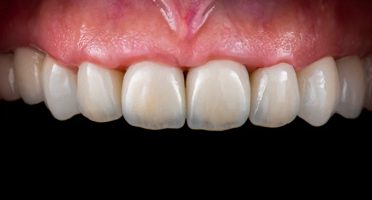
Anterior crowns remain an important component of fixed prosthodontics, combining aesthetic requirements with mechanical function in an area of the mouth where appearance is of primary concern. The successful use of anterior crowns requires a detailed understanding of material selection, biological considerations, technical procedures, and common clinical difficulties.
Table of Contents
TogglePre-operative Considerations
Oral Hygiene and Disease Control
Preparation for any crown should be deferred until the patient demonstrates adequate oral hygiene. Healthy gingival tissues are essential to achieving accurate impressions and predictable placement of preparation margins. Gingival inflammation can obscure the true gingival contour, bleed excessively, and complicate both tooth preparation and impression procedures. Furthermore, periodontal disease and active caries must be stabilised before a definitive crown is considered.
Assessment of Vitality and Radiographic Examination
Prior to preparing a tooth for an anterior crown, its pulpal status must be confirmed. If the tooth is non-vital or displays symptoms or signs of irreversible pulpitis or apical pathology, endodontic treatment should be completed first. A periapical radiograph is indispensable for assessing apical status, supporting bone levels, periodontal ligament width, existing restorations, and the general anatomy of the root.
Study Models, Diagnostic Wax-ups, and Treatment Planning
Study models provide essential three-dimensional information for diagnosis and treatment planning. Diagnostic wax-ups—particularly useful for less experienced practitioners—allow prediction of the final aesthetic and functional outcome and help the operator visualise the required tooth reduction. They can also be used to fabricate reduction guides or provisional restorations.
Shade selection must be performed before the tooth dehydrates during preparation. The shade should be assessed under both natural and artificial light sources. Occlusion must also be analysed carefully. The presence of parafunction, deep overbite, or unfavourable occlusal patterns can greatly influence material choice and preparation design.
Types of Anterior Crowns
Porcelain Fused to Metal (PFM) Crowns
PFM crowns combine the strength of a metal substructure with the aesthetic potential of porcelain. Although less aesthetic than modern all-ceramic systems, PFM crowns remain a predictable and durable option, particularly when additional mechanical strength is required.
Preparation Requirements
The typical preparation includes:
- 0.5 mm reduction of the lingual surface, with a chamfer margin
- 1.2–1.5 mm labial reduction, allowing adequate porcelain thickness
- Chamfered shoulder on the labial aspect
- Proximal shoulder blending to chamfer lingually
The porcelain–metal junction should ideally be in non-contact zones to reduce the risk of chipping. In the incisal region, occlusal contacts should preferably be in metal when strength is critical, though this compromises translucency.
PFMs offer excellent longevity but are limited aesthetically by the opacity of the metal substructure and potential gingival margin greying caused by metal collar visibility.
Porcelain Jacket Crowns (PJC)
PJCs were historically the restoration of choice for anterior aesthetics before the introduction of stronger all-ceramic systems. They are constructed entirely of porcelain without a metal core, thus providing superior translucency and natural appearance.
Their main drawback is their lower strength, making them more prone to fracture under high occlusal loads. Nevertheless, they remain a useful reference point in prosthodontic history and may still be indicated in selected low-load aesthetic cases.
Dentine-Bonded Crowns
Dentine-bonded ceramic crowns rely on resin bonding technology to achieve retention and strength. They use etched ceramic on the fitting surface, a dentine bonding agent, and a resin cement to form a micromechanical and chemical bond with the tooth structure.
Advantages
- Minimal labial reduction is required, preserving tooth structure
- Excellent aesthetics, as the crown becomes more optically integrated with the underlying dentine
- Very useful in tooth wear cases or situations where the axial walls are short and conventional retention is lacking
Considerations
Lingual reduction must still be sufficient to allow material strength. Chair-side bonding procedures are more technique-sensitive and time-consuming compared with traditional crown cementation.
All-Ceramic Crowns
Modern all-ceramic materials offer improved strength, fracture resistance, and aesthetics. They are suitable for most anterior restorative needs and have largely replaced PJCs.
Indications
- Cases requiring superior aesthetics
- Teeth with existing large restorations
- Situations where dentine-bonded systems are unsuitable
- Cases where masking of discoloured substrates is required (depending on material)
Examples of Materials
- Lithium disilicate (e.g., IPS e.max) – high translucency, excellent aesthetics, resin-bonded
- Glass-ceramics (e.g., IPS Empress) – good aesthetics, etchable
- Zirconia-based ceramics (e.g., Lava, Procera, In-Ceram, Zircon) – very strong but more opaque; cannot be etched; cemented with RMGIC or resin cement
- Alumina-based ceramics – opaque and strong, suitable for masking dark substrates
The choice of ceramic depends on the aesthetic and functional demands of the case.
Principles of Tooth Preparation
Successful crown preparation aims to provide adequate space for restorative materials while preserving tooth structure and maintaining pulpal and periodontal health.
Amount of Reduction
- Labial: ~1.0 mm (two-plane reduction following natural contours)
- Palatal: Supragingival chamfer, maintaining 0.8–1.0 mm clearance with opposing teeth
- Incisal: 1.5–2.0 mm reduction for strength and translucency
- Proximal: Tapered chamfer bur, 6° taper, converging lingually
Taper
A total convergence angle of ~6° between opposing walls optimises retention and resistance form.
Finishing
Line angles should be rounded to reduce stress concentration within the ceramic and improve seating of the restoration.
Detailed Preparation Steps
1. Shade Selection
Shade is assessed in both natural and artificial light with the tooth hydrated. The shade guide should be held at arm’s length and compared rapidly to avoid visual fatigue.
2. Interproximal Reduction
A long tapered chamfer bur is used to break the proximal contacts, creating space for the crown material while preserving gingival health. Care must be taken to avoid gouging.
3. Labial Reduction
Three depth-cut grooves are placed to control reduction. The labial surface is reduced in two planes:
- cervical third following the convexity of the tooth
- incisal two-thirds following the natural inclination
Subgingival extension should not exceed 0.5 mm unless required for aesthetic reasons or to hide margins.
4. Lingual Reduction
Initial grooves guide the reduction. Supragingival margins are preferred where possible. A flame-shaped bur creates a smooth chamfer, ensuring that the palatal clearance allows proper occlusal function.
5. Incisal Reduction
An incisal reduction of 1.5–2.0 mm is essential to provide adequate strength and translucency. This also allows the technician space to reproduce incisal edge morphology and internal effects.
6. Finishing
After completing preparation, the margins are refined, undercuts are removed, and any sharp angles are smoothed.
Temporary Crowns
Temporary (provisional) crowns protect the prepared tooth, maintain aesthetics, prevent hypersensitivity, and ensure positional stability. They can be fabricated using a pre-operative impression or matrix, or from a diagnostic wax-up.
Temporaries should:
- Fit accurately
- Maintain occlusal contacts
- Have acceptable aesthetics
- Be cemented with non-eugenol temporary cement to avoid interference with resin bonding
If time is limited, impressions for the final restoration may be deferred, but fabrication of the temporary crown should never be omitted.
Impression Techniques
An accurate impression is essential for a well-fitting crown.
Materials
- Elastomeric materials (e.g., polyvinyl siloxane, polyether) are preferred for their accuracy.
- Alginate can be used for the opposing arch for cost-effectiveness.
- Digital impressions may reduce errors and improve efficiency.
Soft Tissue Management
Retraction cords or retraction pastes may be required to expose the margins. Hemostasis must be achieved prior to impression.
Optical impressions eliminate the need for impression materials, offer increased patient comfort, and may shorten laboratory turnaround time.
Crown Fitting and Cementation
Evaluation
When fitting the definitive crown:
- Ensure optimal isolation
- Remove temporary crown and cement
- Clean the abutment thoroughly
- Check marginal integrity with an explorer
- Assess proximal contacts using floss
- Verify occlusion in static and dynamic movements
Cementation
- Resin cement: used for etchable ceramics and dentine-bonded crowns
- Resin-modified glass ionomer cement (RMGIC): used for zirconia and alumina-based ceramics
If adjustments are needed, porcelain polishing wheels should be used to restore a smooth, glaze-like finish to avoid plaque accumulation and wear on opposing teeth.
Before cementation, ensure the patient is satisfied with shade and appearance.
Common Problems and Their Management
1. Pulp Exposure Risk
If caries removal or reduction places the pulp at risk, it may be appropriate to delay crowning and provide a veneer-style or temporary restoration. Definitive treatment can be reconsidered once pulpal status is certain.
2. Crown Not Seating
Possible causes include:
- Temporary cement remnants
- Tight proximal contacts
- Distorted impression due to undercuts or material issues
- Over-trimmed die leading to an oversized internal crown surface
Each factor should be systematically investigated and corrected.
3. Visible Core Material
If the underlying core material shows through the ceramic, additional reduction or use of a more opaque core ceramic may be required.
4. Incorrect Shade
Surface characterisation by the technician may correct minor mismatches. Otherwise, the crown must be remade with an accurate shade selection process.
Removing Old Crowns
Old crowns may need to be removed due to recurrent caries, poor fit, aesthetic concerns, or failure of underlying restorations.
Principles
Protect the patient’s airway with gauze or throat packs
Use a crown remover where possible to retrieve the restoration intact
If destruction is necessary:
Cut a longitudinal groove through the crown on labial, palatal, and occlusal surfaces
Avoid damaging underlying tooth structure
Insert a flat plastic instrument into the groove and twist to separate the crown
Safe and controlled removal is essential to avoid iatrogenic damage.
Conclusion
Anterior crowns represent a sophisticated balance between aesthetic demands and mechanical strength. Success in their placement requires careful treatment planning, understanding of material properties, precise tooth preparation, and skilful execution of both provisional and definitive stages. Modern ceramics offer clinicians excellent opportunities to provide natural-appearing restorations with long-term durability, but each material has its own indications and requirements.
Attention to detail—from the initial diagnosis through to final cementation—ensures optimal outcomes for both clinician and patient. Effective management of complications and understanding of crown removal techniques further enhance the clinician’s ability to deliver predictable, high-quality care.
References
- Rosenstiel, S. F., Land, M. F., & Fujimoto, J. (2015). Contemporary fixed prosthodontics (5th ed.). St. Louis: Mosby Elsevier.
- Shillingburg, H. T., Hobo, S., Whitsett, L. D., Jacobi, R., & Brackett, S. E. (2012). Fundamentals of fixed prosthodontics (4th ed.). Chicago: Quintessence Publishing.
- Goodacre, C. J., Campagni, W. V., & Aquilino, S. A. (2001). Tooth preparations for complete crowns: An art form based on scientific principles. Journal of Prosthetic Dentistry, 85(4), 363–376.
- Donovan, T. E., Cho, G. C., & Lee, S. M. (2014). Single-unit anterior all-ceramic crowns: A literature review. Journal of Esthetic and Restorative Dentistry, 26(2), 82–94.
- Kelly, J. R., & Benetti, P. (2011). Ceramic materials in dentistry: Historical evolution and current practice. Australian Dental Journal, 56(s1), 84–96.
- McLean, J. W. (1991). Evolution of dental ceramics in the twentieth century. Journal of Prosthetic Dentistry, 66(4), 512–522.
- Peumans, M., Van Meerbeek, B., Lambrechts, P., & Vanherle, G. (2000). Porcelain veneers: A review of the literature. Journal of Dentistry, 28(3), 163–177.
- Pjetursson, B. E., & Sailer, I. (2007). A systematic review of the survival and complication rates of all-ceramic and metal–ceramic reconstructions. Clinical Oral Implants Research, 18(s3), 73–85.
- Burke, F. J. T. (2012). Tooth preparation for crowns and bridges. BDJ Team, 213(7), 315–320.
- Hill, E. E. (2007). Dental cements for definitive luting: A review and practical clinical considerations. Dental Clinics of North America, 51(3), 643–658.
- Magne, P., & Belser, U. (2003). Bonded porcelain restorations in the anterior dentition: A biomimetic approach. Chicago: Quintessence Publishing.
- Christensen, G. J. (2007). Porcelain-fused-to-metal versus zirconia-based ceramic restorations. Journal of the American Dental Association, 138(9), 1263–1265.
- McLaren, E. A., & Whiteman, Y. Y. (2010). Ceramics in dentistry—Part I: Classification and selection. Compendium of Continuing Education in Dentistry, 31(9), 666–668.
- Sailer, I., Fehér, A., Filser, F., Gauckler, L. J., Lüthy, H., & Hämmerle, C. H. (2007). Prospective clinical study of zirconia posterior fixed partial dentures: 3-year follow-up. International Journal of Prosthodontics, 20(4), 383–388.