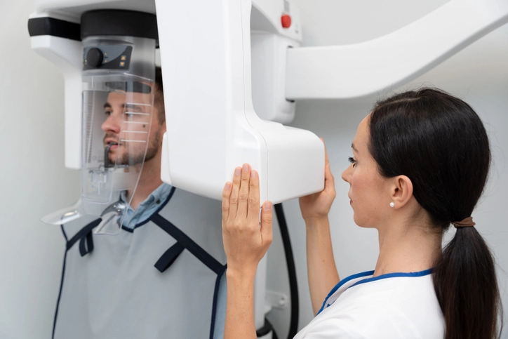
Medical imaging has transformed healthcare, enabling practitioners to visualize internal structures of the human body with remarkable precision. From its early beginnings with X-rays to the sophisticated three-dimensional reconstructions of today, imaging has become indispensable in diagnosis, treatment planning, and monitoring of diseases. In dentistry, maxillofacial surgery, oncology, and general medicine, advanced imaging plays a crucial role in improving patient outcomes, reducing invasive procedures, and guiding surgical interventions.
Table of Contents
ToggleComputed Tomography (CT)
Principle and Technology
Computed tomography (CT) represents one of the most revolutionary innovations in medical imaging. Unlike conventional radiographs, which provide two-dimensional shadow images, CT creates cross-sectional images of the body by rotating a narrow X-ray beam around a patient. As the X-rays pass through tissues, detectors measure the attenuation, and sophisticated computer algorithms reconstruct these signals into slices, which can then be stacked to form three-dimensional images.
A critical aspect of CT imaging is the measurement of Hounsfield Units (HU), which quantify tissue density relative to water. Air, for example, measures around -1000 HU, while bone measures upwards of +1000 HU. This allows clinicians to differentiate between soft tissues, fluids, bone, and pathological changes with high accuracy.
Clinical Applications
CT is invaluable in trauma cases, where rapid assessment of fractures, hemorrhages, or internal injuries is required. In dentistry and maxillofacial surgery, CT scans help in evaluating complex fractures, planning orthognathic surgery, and assessing extensive tumors. In oncology, CT is used for staging cancers, monitoring treatment response, and detecting metastases.
Advantages
- Provides detailed anatomical visualization.
- Enables 3D reconstruction for surgical planning.
- Rapid image acquisition, making it suitable for emergencies.
Limitations
- Relatively high radiation dose compared to plain radiographs.
- Limited soft tissue contrast compared to MRI.
- Expensive equipment and infrastructure requirements.
Future innovations in CT, including dose-reduction algorithms and faster scanning technologies, are making it safer and more accessible.
Cone Beam Computed Tomography (CBCT)
Principle and Development
CBCT emerged as a modification of CT tailored to dentistry and maxillofacial imaging. Instead of using fan-shaped X-ray beams, CBCT employs cone-shaped beams that cover the entire region of interest in a single rotation. The result is a three-dimensional reconstruction of craniofacial structures with lower radiation doses compared to conventional CT.
Clinical Applications
CBCT is particularly useful in:
- Implantology: assessing bone volume, density, and anatomical landmarks before implant placement.
- Endodontics: detecting root canal morphology, periapical lesions, and resorptive defects.
- Orthodontics: evaluating skeletal discrepancies and airway analysis.
- Surgical planning: assisting with impacted tooth removal, fracture management, and temporomandibular joint (TMJ) imaging.
Advantages
- Lower radiation exposure than conventional CT.
- High spatial resolution tailored to dentomaxillofacial structures.
- Relatively quick and accessible.
Limitations
- Not standardized across machines, making HU measurements less reliable.
- Susceptible to artefacts from patient movement and metallic restorations.
- Limited soft tissue contrast.
CBCT continues to revolutionize dentistry, and with ongoing improvements in resolution and dose optimization, its role is expected to expand further.
Magnetic Resonance Imaging (MRI)
Principle and Physics
MRI represents a fundamentally different approach to imaging compared to CT. Instead of using ionizing radiation, MRI leverages strong magnetic fields and radiofrequency waves to exploit the behavior of hydrogen atoms in tissues. Since hydrogen is abundant in the human body, particularly in water and fat, MRI is highly effective in producing detailed soft tissue images.
When placed in a magnetic field, hydrogen protons align in a predictable manner. Radiofrequency pulses disturb this alignment, and as the protons “relax” back, they emit signals captured by detectors. These signals vary depending on tissue properties, creating two main types of images: T1-weighted (better for anatomy) and T2-weighted (better for pathology and fluids).
Clinical Applications
MRI is the gold standard for imaging soft tissues, including the brain, spinal cord, muscles, joints, and internal organs. In dentistry and maxillofacial practice, MRI is invaluable for:
- Assessing temporomandibular joint (TMJ) disorders.
- Imaging soft tissue lesions and tumors.
- Evaluating inflammatory changes in soft tissues.
Advantages
- No ionizing radiation, making it safe for repeated use.
- Superior soft tissue contrast compared to CT and CBCT.
- Useful in differentiating tissue types and fluid states.
Limitations
- Long scan times, making it sensitive to patient movement.
- High cost and limited availability in some settings.
- Contraindicated in patients with pacemakers, ferromagnetic implants, or severe claustrophobia.
Advancements in MRI include functional imaging (fMRI), diffusion-weighted imaging (DWI), and faster sequences, expanding its diagnostic potential.
Digital Imaging
Concept and Development
Digital imaging has replaced conventional film-based radiology in many practices. Instead of using X-ray films, digital systems employ electronic sensors to capture and display images instantly. These images can be manipulated, magnified, and enhanced for diagnostic accuracy.
Clinical Applications
Digital imaging is now routine in dental radiographs (intraoral, panoramic, and cephalometric), chest X-rays, and many other diagnostic fields. Its integration with electronic health records facilitates better communication between healthcare providers.
Advantages
- Lower radiation doses compared to film radiography.
- Instant image availability, reducing patient wait times.
- Easy image storage, retrieval, and sharing.
- Ability to adjust contrast and brightness for improved interpretation.
Limitations
- Lower resolution compared to traditional film in some cases.
- High initial cost of equipment.
- Requires proper calibration and quality assurance.
Despite these limitations, digital imaging has become the new standard due to its efficiency, cost-effectiveness, and environmental sustainability.
Ultrasound (US)
Principle
Ultrasound uses high-frequency sound waves (1–20 MHz) transmitted into the body through a transducer containing piezoelectric crystals. As sound waves encounter tissues, they are reflected, refracted, or absorbed depending on tissue density and interfaces. These echoes are processed to form real-time images.
Clinical Applications
- Dentistry and maxillofacial surgery: evaluating cysts, abscesses, and salivary gland pathologies.
- Medicine: abdominal imaging, obstetrics, cardiology, vascular studies, and musculoskeletal assessments.
- Doppler ultrasound: assessing blood flow, vascular patency, and lesion vascularity.
Advantages
- Safe, non-invasive, and free from ionizing radiation.
- Portable and relatively inexpensive.
- Provides real-time imaging.
Limitations
- Operator-dependent and requires skill for accurate interpretation.
- Limited penetration through bone or air-filled cavities.
- Image quality can be affected by obesity or deep structures.
Emerging developments like elastography and contrast-enhanced ultrasound are expanding its diagnostic capabilities.
Sialography
Principle
Sialography involves injecting a radiopaque contrast medium into the salivary ducts, followed by imaging (X-ray, CT, or digital methods). It outlines the architecture of salivary glands and detects obstructions, strictures, or stones.
Clinical Applications
- Diagnosis of sialolithiasis (salivary stones).
- Evaluation of chronic sialadenitis (inflammation).
- Assessing ductal abnormalities.
Advantages
- Provides detailed ductal anatomy.
- Helpful in differentiating obstructive from inflammatory conditions.
Limitations
- Invasive and uncomfortable for patients.
- Contraindicated in acute infections and patients allergic to contrast media.
- Risk of anaphylaxis to iodine-based contrast.
Newer non-invasive alternatives like sialoendoscopy and MRI sialography are reducing reliance on traditional sialography.
Arthrography
Principle
Arthrography involves injecting contrast media into a joint cavity, followed by fluoroscopic imaging. This technique allows visualization of joint spaces, articular surfaces, and intra-articular structures.
Clinical Applications
- Imaging of temporomandibular joint (TMJ) disorders.
- Assessing meniscal tears, ligament injuries, and joint effusions.
- Combined with MRI (MR arthrography) for enhanced accuracy.
Advantages
- Useful for visualizing intra-articular abnormalities.
- Provides real-time dynamic imaging.
Limitations
- Technically challenging and invasive.
- Risk of infection, bleeding, or allergic reaction.
- Interpretation may be difficult compared to modern MRI.
Although increasingly replaced by MRI, arthrography remains useful in selected cases.
Positron Emission Tomography (PET)
Principle
PET relies on the detection of positrons (beta particles) emitted from radiotracers introduced into the body. The most commonly used tracer is fluorodeoxyglucose (FDG), which highlights areas of increased metabolic activity. Since malignant cells often have higher glucose metabolism, PET scans are highly effective in oncology.
Clinical Applications
- Cancer detection, staging, and treatment monitoring.
- Differentiating between recurrent tumor and scar tissue.
- Evaluating metabolic activity in neurological and cardiac disorders.
Advantages
- Functional imaging that detects disease before structural changes occur.
- High sensitivity in detecting malignancy.
- Can be combined with CT (PET/CT) or MRI (PET/MRI) for anatomical correlation.
Limitations
- Expensive and not widely available.
- Limited resolution compared to CT or MRI.
- Exposure to radiation through radiotracers.
PET continues to be a cornerstone in oncology and research into novel tracers is expanding its utility to neurodegenerative and inflammatory diseases.
Conclusion
Advanced imaging techniques have revolutionized diagnostics in both medicine and dentistry. From the anatomical precision of CT and CBCT to the soft tissue detail of MRI and the functional insights of PET, each modality offers unique advantages. Digital imaging has streamlined workflows, while ultrasound provides a safe, real-time tool. Although invasive, techniques like sialography and arthrography remain relevant in select contexts.
The future of imaging lies in hybrid technologies (such as PET/MRI), artificial intelligence for automated interpretation, and continued efforts to minimize radiation exposure. As technology advances, the integration of these modalities will further enhance patient care, making imaging not just a diagnostic tool but an integral part of personalized and predictive medicine.
References
- White, S. C., & Pharoah, M. J. (2019). Oral Radiology: Principles and Interpretation (8th ed.). St. Louis: Elsevier.
→ Comprehensive coverage of CBCT, CT, MRI, ultrasound, and sialography in dental and maxillofacial applications. - Langland, O. E., Langlais, R. P., & Preece, J. W. (2016). Principles of Dental Imaging (2nd ed.). Wolters Kluwer Health.
→ Explains fundamental imaging techniques, radiation safety, and digital radiography in dentistry. - Hendee, W. R., & Ritenour, E. R. (2011). Medical Imaging Physics (4th ed.). Wiley-Liss.
→ Standard reference for understanding CT, MRI, and ultrasound physics and principles. - Bushberg, J. T., Seibert, J. A., Leidholdt, E. M., & Boone, J. M. (2020). The Essential Physics of Medical Imaging (4th ed.). Wolters Kluwer Health.
→ Core text detailing CT, MRI, and PET physics, applications, and safety. - Ludlow, J. B., Timothy, R., Walker, C., Hunter, R., Benavides, E., Samuelson, D. B., & Scheske, M. J. (2015). Effective dose of dental CBCT—a meta-analysis. Dentomaxillofacial Radiology, 44(1):20140197.
→ Provides data comparing radiation doses in CBCT and conventional CT. - Boeddinghaus, R., & Whyte, A. (2008). Current concepts in maxillofacial imaging. European Journal of Radiology, 66(3), 396–418.
→ Discusses modern imaging modalities used in maxillofacial diagnosis and treatment planning. - Scarfe, W. C., & Farman, A. G. (2008). What is cone-beam CT and how does it work? Dental Clinics of North America, 52(4), 707–730.
→ Authoritative source on CBCT technology, reconstruction, and diagnostic potential. - Casselman, J. W., et al. (2000). Imaging of the temporomandibular joint using MRI and CT. European Journal of Radiology, 34(2), 102–118.
→ Key reference for TMJ imaging with MRI and CT. - National Radiological Protection Board (NRPB). (2001). Guidelines on radiation dose and safety in diagnostic imaging.
→ Provides recommended dose limits and safety standards for CT and CBCT. - Weber, A. L., & Randolph, G. (1998). Sialography and imaging of the salivary glands. Radiologic Clinics of North America, 36(5), 941–961.
→ Classic paper on techniques and interpretation of sialography. - Townsend, D. W. (2008). Multimodality imaging of structure and function. Physics in Medicine and Biology, 53(4), R1–R39.
→ Describes the integration of PET/CT and PET/MRI in oncology and neuroscience. - World Health Organization (WHO). (2022). Radiation protection in diagnostic and interventional radiology. WHO Technical Report Series.
→ International safety guidelines for radiation exposure and patient protection. - Rubin, G. D. (2014). Computed tomography: Revolutionizing the practice of medicine for 40 years. Radiology, 273(2S), S45–S74.
→ Overview of CT evolution, technology, and clinical impact.

