
The process of tooth eruption and exfoliation represents one of the most intricate and coordinated developmental phenomena in human growth. Eruption refers to the movement of a tooth from its developmental position within the jawbones into its functional position in the oral cavity, while exfoliation denotes the natural shedding of primary teeth to make way for their permanent successors.
Disruptions in these processes—whether through premature eruption, delayed eruption, failure to erupt, or premature exfoliation—may serve as early indicators of local or systemic pathology and can have lasting implications for occlusion, mastication, and facial development.
Table of Contents
ToggleNatal and Neonatal Teeth
Definition and Incidence
- Natal teeth are those present at birth.
- Neonatal teeth erupt within the first 30 days after birth.
These are most often part of the normal primary dentition, though occasionally they are supernumerary.
The incidence ranges between 1:2,000 and 1:3,500 live births. The mandibular central incisors are the most commonly affected.
Etiology
The exact cause is uncertain, but several hypotheses exist:
- Superficial position of the tooth germ close to the alveolar crest.
- Endocrine factors, including increased activity of the thyroid or pituitary glands.
- Nutritional deficiency, particularly hypovitaminosis.
- Genetic predisposition or syndromic associations (e.g., Ellis–van Creveld syndrome, Hallermann–Streiff syndrome).
Clinical Features
- Teeth may appear immature and hypoplastic, with poorly formed roots.
- They are often mobile, posing a risk of aspiration.
- The mother may experience nipple trauma during breastfeeding.
- Surrounding soft tissues may become ulcerated (notably Riga–Fede disease due to repetitive trauma).
Management
Retention is preferred if the tooth is stable and does not interfere with feeding.
Extraction is indicated when:
The tooth is supernumerary.
There is marked mobility (risk of aspiration).
Feeding difficulties or ulcerations occur.
Extraction should be carried out with minimal or no local anesthesia due to incomplete root formation, and the site may be gently curetted to prevent residual odontogenic tissue persistence.
Teething
Definition and Background
Teething refers to the eruption of primary teeth through the gingiva and typically begins at 6–8 months of age, continuing until about 2½–3 years.
Though a normal physiological process, it has long been associated with a range of systemic symptoms.
Clinical Manifestations
Common signs include:
- Localized gingival irritation, itching, or erythema.
- Increased salivation.
- Desire to bite or chew firm objects.
- Mild fever, restlessness, and sleep disturbances in some cases.
While many caregivers attribute systemic symptoms such as diarrhea or high fever to teething, research indicates that teething itself does not cause significant systemic illness.
Management
- Reassurance to parents is essential.
- Teething rings or cooled (not frozen) objects may provide soothing relief.
- Topical teething gels containing mild anesthetic, antiseptic, or anti-inflammatory agents can be used sparingly.
- Avoid excessive use of systemic medications.
- Persistent or severe symptoms warrant evaluation for alternative causes such as infection or nutritional deficiencies.
Eruption Cysts
Definition
An eruption cyst is a soft tissue cyst that develops over an erupting tooth, resulting from the accumulation of fluid or blood within the follicular space.
Clinical Features
- Appears as a bluish, translucent, dome-shaped swelling overlying an erupting tooth.
- Commonly occurs in the deciduous molar or permanent incisor regions.
- The lesion is painless, unless infected.
- The presence of blood imparts a bluish or purplish hue (sometimes termed an eruption hematoma).
Management
- Most rupture spontaneously, allowing eruption to continue.
- If eruption is delayed or the cyst causes discomfort, a simple marsupialization or surgical excision of the cystic roof facilitates normal eruption.
- No extraction or extensive surgery is necessary.
Failure or Delayed Eruption
Definition
Failure or delayed eruption refers to the absence of tooth emergence into the oral cavity beyond the expected age range, considering the individual’s developmental rather than chronological age.
Etiology
A. General Causes
- Hereditary gingival fibromatosis
- Down syndrome
- Gardner syndrome
- Hypothyroidism
- Cleidocranial dysostosis
- Rickets
B. Local Causes
- Congenital absence of the tooth germ (anodontia, hypodontia).
- Crowding in the dental arch, obstructing eruption path.
- Retention of primary tooth due to failure of root resorption.
- Supernumerary teeth, especially mesiodens, blocking eruption.
- Crown or root dilaceration due to trauma to primary predecessors.
- Abnormal position of crypt or ectopic eruption path.
- Ankylosis of the tooth root with alveolar bone.
- Alveolar bone sclerosis or fibrotic tissue impeding eruption.
Clinical Features
- Delayed appearance of one or more teeth compared to contralateral or sequential teeth.
- Possible asymmetry in eruption (difference >6 months between contralateral teeth).
- Retained primary teeth or missing teeth in the arch.
- Possible localized swelling if cystic obstruction exists.
Diagnostic Evaluation
- Radiographic examination (OPG, periapical, or occlusal radiographs) to assess tooth position, presence of obstruction, or pathology.
- Endocrine and systemic assessment if generalized delay is noted.
Management
- Observation in mild developmental delays.
- Extraction of retained primary teeth or supernumerary teeth obstructing eruption.
- Orthodontic intervention (e.g., surgical exposure and traction) for impacted permanent teeth.
- Medical treatment for systemic disorders affecting eruption (e.g., hormone replacement in hypothyroidism).
Infraoccluded (Ankylosed) Primary Molars
Definition
Infraocclusion (ankylosis) occurs when a primary molar fails to maintain its normal occlusal level relative to adjacent teeth due to fusion between the cementum and alveolar bone.
Etiology
- Localized trauma to the periodontal ligament.
- Genetic predisposition.
- Absence or malformation of the permanent successor, resulting in retention and ankylosis of the primary tooth.
- Disturbance in bone metabolism or infection.
Clinical Features
- The affected tooth lies below the occlusal plane of adjacent teeth.
- Percussion test produces a sharp metallic sound, characteristic of ankylosis.
- Adjacent teeth may tip towards the ankylosed tooth.
- In severe cases, the tooth appears submerged below the gingival level.
- Loss of arch length and occlusal discrepancies may develop.
Radiographic Features
- Absence of a normal periodontal ligament space.
- Loss of root resorption process compared to normal exfoliation pattern.
Management
Observation if the ankylosed tooth exfoliates normally and the permanent successor is erupting.
Extraction if:
There is significant infraocclusion.
The permanent successor fails to erupt.
There is drifting or tilting of adjacent teeth.
Space maintenance is crucial post-extraction to prevent malocclusion.
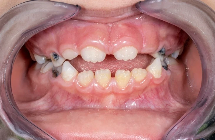
Ectopic Eruption of Permanent First Molars
Definition
An ectopic eruption is the abnormal path of eruption of a tooth, most commonly affecting the maxillary permanent first molars, where the tooth erupts mesially and becomes locked under the distal aspect of the second primary molar.
Epidemiology
Occurs in 2–5% of children, with a higher incidence in the maxilla.
The condition may be self-correcting (jump type) or non-correcting (hold type).
Etiology
- Abnormal eruption angulation (mesial tilt of the molar).
- Lack of space in the arch.
- Early eruption of permanent molars.
- Abnormally large crown size of the permanent molar.
- Developmental anomalies or crowding in the posterior segment.
Clinical Features
- Delayed eruption of the permanent molar.
- Resorption of the distal root of the adjacent primary second molar.
- Sometimes presents as pain or localized swelling.
- May lead to early loss of the second primary molar, reducing arch length and causing crowding.
Radiographic Findings
- Mesial inclination of the permanent molar crown impinging on the distal aspect of the primary molar.
- Resorption of the primary molar root seen on bitewing or periapical radiographs.
Management
- Observation: In mild cases (jump type), spontaneous correction occurs within 4–6 months.
- Separation appliances: Brass wire separators, orthodontic separators, or elastic modules can help guide eruption.
- Surgical or orthodontic intervention: In severe or non-correcting cases, extraction of the primary molar may be necessary once the permanent molar is stable in position.
- Space maintenance to prevent mesial drift is mandatory after extraction.
Premature Exfoliation
Definition
Premature exfoliation refers to the loss of primary teeth earlier than their normal exfoliation period, not due to natural resorption by permanent successors.
Etiology
A. Local Causes
- Caries leading to extraction or loss due to infection.
- Trauma resulting in avulsion or subluxation.
B. Systemic Causes
Periodontal and bone diseases:
Prepubertal periodontitis.
Hypophosphatasia (low alkaline phosphatase activity).
Langerhans cell histiocytosis.
Papillon–Lefèvre syndrome.
Hematological disorders:
Cyclic neutropenia.
Leukemia.
Agranulocytosis.
Nutritional and metabolic causes:
Vitamin D–resistant rickets.
Hypophosphatemia.
Clinical Features
- Early tooth loss, often multiple.
- Associated gingival inflammation or bone loss.
- Increased tooth mobility without apparent caries.
- Spacing and drifting of remaining teeth.
Management
- Identify and treat the underlying cause (hematological or metabolic).
- Space maintenance to preserve arch integrity until eruption of permanent successors.
- Oral hygiene reinforcement and regular monitoring for bone resorption.
- In systemic disease, coordination with pediatricians and hematologists is essential.
Clinical and Diagnostic Considerations
Assessment of Eruption Disturbances
History taking:
Family history of delayed eruption or missing teeth.
Systemic illness or syndromes.
Past trauma to primary teeth.
Clinical examination:
Inspection for asymmetry, retained teeth, infraocclusion, or abnormal sequence.
Palpation for bulging or cystic lesions over erupting teeth.
Radiographic examination:
OPG (orthopantomogram) for general assessment.
Periapical or occlusal radiographs for localized details.
CBCT in complex impactions.
Laboratory tests:
Thyroid function, serum calcium, phosphate, and alkaline phosphatase for metabolic causes.
Clinical Significance
- Disturbances in eruption and exfoliation can affect occlusion, masticatory efficiency, speech development, and facial aesthetics.
- Early detection allows for timely intervention and prevention of complex orthodontic problems.
- Persistent retained teeth or infraoccluded molars can lead to arch length discrepancies, impaction, or pathological root resorption.
- Abnormal premature exfoliation may signal serious systemic disease, warranting multidisciplinary evaluation.
Preventive and Therapeutic Strategies
- Regular dental check-ups during childhood to monitor eruption sequence.
- Education of parents to identify early signs of abnormal eruption.
- Radiographic screening in cases of delayed or asymmetric eruption.
- Interceptive orthodontic measures such as extraction of primary teeth, use of separators, or space maintainers.
- Systemic management of underlying endocrine or metabolic disorders.
- Referral to pediatric specialists for systemic disease manifestations like neutropenia or rickets.
Conclusion
Abnormalities of tooth eruption and exfoliation encompass a broad spectrum of local and systemic conditions that can profoundly influence the developing dentition. Early recognition and accurate diagnosis are essential for preventing long-term sequelae such as malocclusion, functional impairment, and aesthetic concerns.
A thorough understanding of normal eruption patterns, combined with vigilant clinical and radiographic assessment, enables the clinician to distinguish between physiological variation and true pathological delay or failure.
Ultimately, successful management requires a multidisciplinary approach, involving pediatric dentists, orthodontists, pediatricians, and, when necessary, medical specialists in endocrinology and hematology.
References
- Cameron, A. C., & Widmer, R. P. (2021). Handbook of Pediatric Dentistry (5th ed.). Elsevier.
→ Comprehensive reference for normal and abnormal tooth eruption patterns, teething, and eruption cysts. - Wright, G. Z., Kupietzky, A., & Welbury, R. R. (2014). Behavior Management in Dentistry for Children (2nd ed.). Wiley-Blackwell.
→ Discusses behavioral considerations during teething and early childhood oral conditions. - McDonald, R. E., Avery, D. R., & Dean, J. A. (2016). McDonald and Avery’s Dentistry for the Child and Adolescent (10th ed.). Elsevier.
→ Authoritative source on pediatric oral pathology, tooth eruption sequences, and exfoliation abnormalities. - Poulton, D. R., & Robinson, P. G. (2002). “Eruption disturbances in the permanent dentition: their etiology, diagnosis and management.” British Dental Journal, 193(7), 387–393.
→ Peer-reviewed journal article providing a clinical overview of eruption disturbances. - Andreasen, J. O., & Andreasen, F. M. (2018). Textbook and Color Atlas of Traumatic Injuries to the Teeth (5th ed.). Wiley-Blackwell.
→ Reference for traumatic causes of delayed or abnormal eruption due to primary tooth injury. - Proffit, W. R., Fields, H. W., Larson, B., & Sarver, D. M. (2018). Contemporary Orthodontics (6th ed.). Elsevier.
→ Details on the sequence and timing of eruption in relation to orthodontic development. - Neville, B. W., Damm, D. D., Allen, C. M., & Chi, A. C. (2015). Oral and Maxillofacial Pathology (4th ed.). Elsevier.
→ Pathological insights into eruption cysts, ankylosis, and systemic causes of eruption failure. - Suri, L., Gagari, E., & Vastardis, H. (2004). “Delayed tooth eruption: Pathogenesis, diagnosis, and treatment. A literature review.” American Journal of Orthodontics and Dentofacial Orthopedics, 126(4), 432–445.
→ Highly cited review of delayed eruption and its multifactorial causes. - Almonaitiene, R., Balciuniene, I., & Tutkuviene, J. (2010). “Factors influencing permanent teeth eruption. Part one – General factors.” Stomatologija, 12(3), 67–72.
→ Scientific discussion of general and systemic factors influencing eruption timing. - American Academy of Pediatric Dentistry (AAPD). (2023). Guideline on Management of the Developing Dentition and Occlusion in Pediatric Dentistry.
→ Clinical guidelines outlining management of eruption disturbances and preventive measures.